
CLEAR Technology
- 27 May 2026
In News:
Researchers at the Jawaharlal Nehru Centre for Advanced Scientific Research, Bengaluru, have developed an innovative protein imaging platform called CLEAR (Cleavable Light-Erased Antibody Reporter). The technology enables scientists to visualize and map a large number of proteins within the same biological sample using a single fluorescent marker, overcoming major limitations of conventional imaging techniques.
Why is Protein Imaging Important?
Proteins are the functional molecules that regulate virtually every biological process within cells. Understanding where proteins are located, how they interact, and how their distribution changes during disease is crucial for deciphering cellular behavior.
In diseases such as cancer, the spatial arrangement of proteins within tissues often determines how tumors grow, evade immunity, and respond to treatment. Therefore, high-resolution protein mapping has become an essential tool in modern biomedical research.
What is CLEAR Technology?
CLEAR (Cleavable Light-Erased Antibody Reporter) is a next-generation multiplex spatial protein imaging platform designed to visualize numerous proteins within the same tissue or cell sample while preserving their spatial organization.
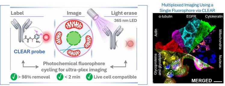
Unlike conventional imaging methods that require multiple fluorescent colours for different proteins, CLEAR repeatedly uses a single fluorescent channel through a unique "erase-and-rewrite" mechanism. This allows scientists to build highly detailed molecular maps without being constrained by the limited number of fluorescent colours detectable by microscopes.
How Does CLEAR Work?
The technology is based on a cyclic imaging process.
Initially, specialized antibodies linked to fluorescent markers through light-sensitive chemical bonds are introduced into a tissue sample. These antibodies bind to specific target proteins, and the sample is imaged under a microscope.
Once imaging is completed, the fluorescent signal is removed by exposing the sample to a gentle pulse of 365 nm LED light. The light breaks the photosensitive linker, effectively erasing the fluorescence without damaging the biological specimen.
The sample then becomes available for a new round of protein labeling and imaging using the same fluorescent channel. By repeating this cycle multiple times, researchers can generate highly detailed spatial maps containing information on numerous proteins within a single specimen.
Key Features
One of the most important strengths of CLEAR is its high-plex multiplexing capability. It allows visualization of a much larger number of proteins than conventional fluorescence microscopy.
The technology is also notable for its live-cell compatibility. Traditional multiplex imaging often requires harsh chemicals or heat treatments to remove fluorescent signals, which can damage tissues and cells. CLEAR relies on gentle light-based erasure, preserving biological integrity.
Another major advantage is its ability to capture the precise spatial distribution of proteins within tissues. Such spatial information is critical for understanding cellular communication, tumor microenvironments, and disease progression.
By repeatedly utilizing a single fluorescent channel, CLEAR also reduces dependence on expensive multi-laser imaging systems, improving cost efficiency and workflow speed.
Significance for Cancer Research
Cancer is not merely a collection of abnormal cells but a complex ecosystem involving tumor cells, immune cells, blood vessels, and surrounding tissues. Understanding this microenvironment requires the simultaneous mapping of multiple proteins.
CLEAR enables researchers to:
- Identify protein interaction networks within tumors.
- Understand mechanisms of cancer progression and metastasis.
- Study immune cell infiltration and tumor immune evasion.
- Discover novel diagnostic biomarkers.
- Improve precision medicine and targeted therapies.
The technology can help researchers develop more personalized treatment approaches based on detailed molecular characteristics of individual tumors.